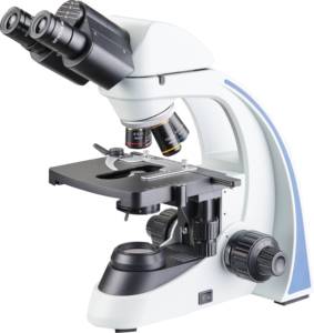
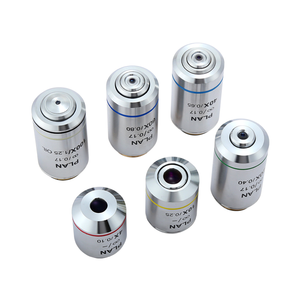
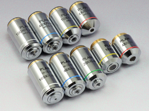
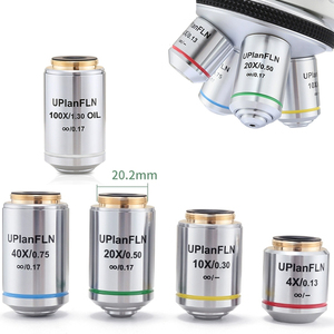
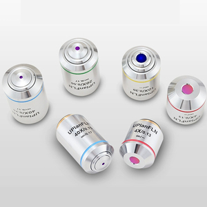
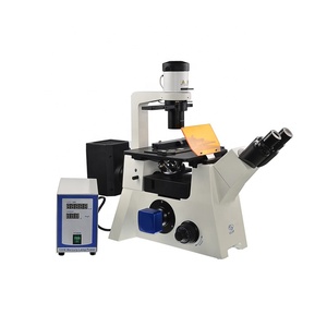
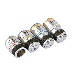
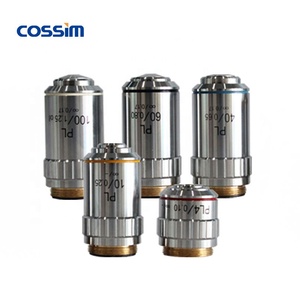
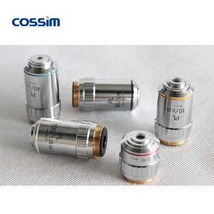
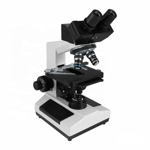
Microscope Objective Types
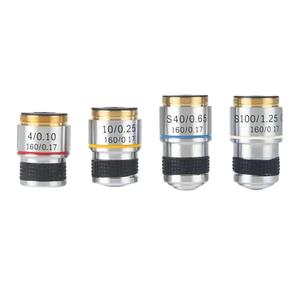
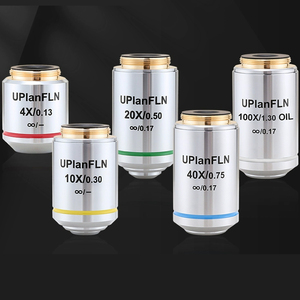
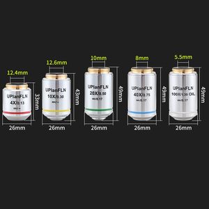
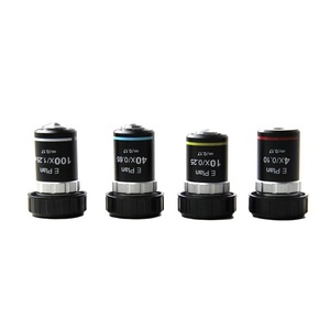
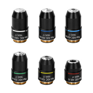
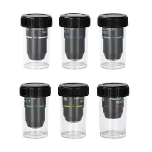
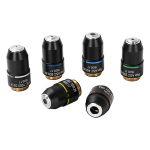
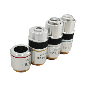
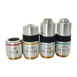
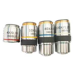
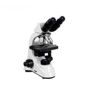
Microscope objective types are important in microscopy as they help in obtaining sharp images at varying resolutions and magnifications for different applications. These objectives, which are mounted on the revolving nosepiece of the microscope, range from low power to high power and are employed based on the specimen being examined. The following are the common types found in majority objective lenses:
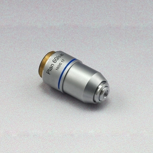
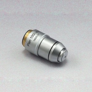
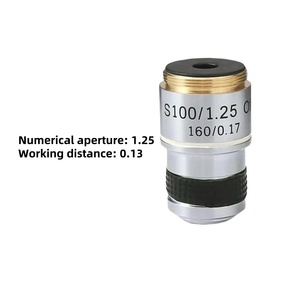
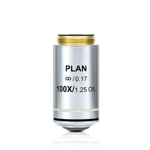
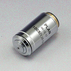
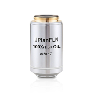
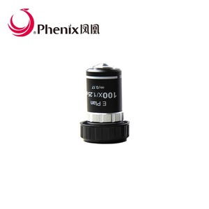
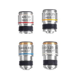
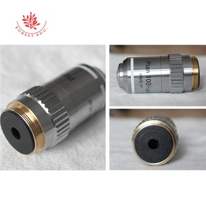
100x Objective Lens
A 100x microscope objective is categorised as a high power lens that provides detailed views of small specimens, such as cells and microorganisms. This objective is mostly used in biological and medical fields where fine structural details are required. Often, this lens is used with a 1x oil diagonal for clear resolution, especially in the evaluation of specimens where precision counts, such as in histology and microbiology.
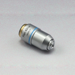
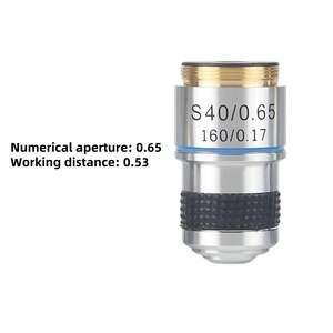
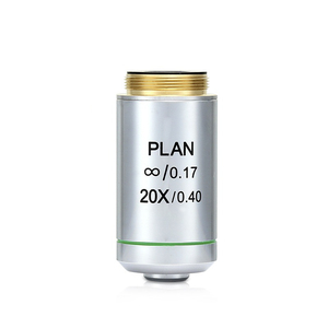
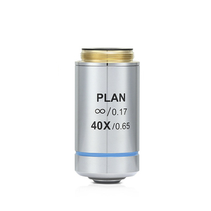
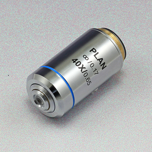
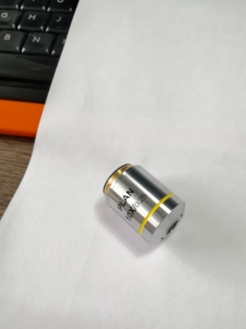
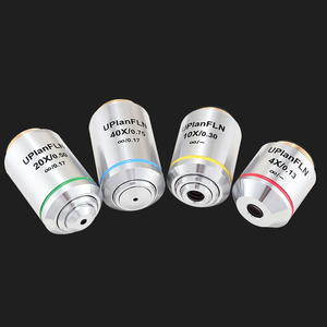
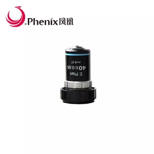
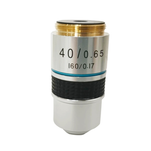
40x Objective Lens
The 40x lens is a high-power lens that provides a detailed view of specimens. Commonly found in lab microscopes, this objective is used in biological and material sciences to provide more information than lower power objectives. It focuses on small areas of specimens with good detail. This lens is well applicable for cell and tissue examinations and is often used together with an oil condenser for enhanced clarity.
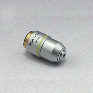
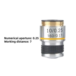
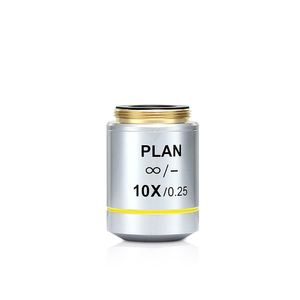
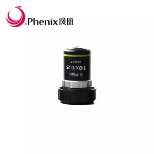
10x Objective Lens
The 10x lens is a medium-power microscope objective lens that provides a moderate magnifying power. This lens strikes a balance between a wide field of view and detailed observation. The 10x lens is suitable for routine lab use and offers flexibility in specimen examination. It is commonly applied in the first stages of specimen observation to provide an overview before switching to higher magnifications.
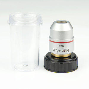
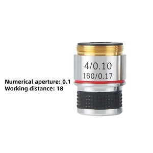
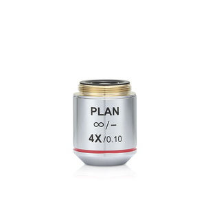
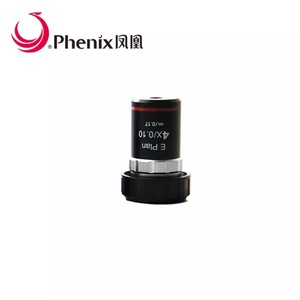
4x Objective Lens
The 4x lens is a low-power objective lens that gives a broad view of specimens. Its low magnification is helpful for scanning large areas or locating specific regions of interest within a sample. It provides a wide field of view that is useful when searching for objects such as cells or tissues. This lens is often used as the starting point in microscopy to quickly find areas that need further detailed examination with higher magnifications.
Features of Microscope Objectives
-
High Magnification
The 100x microscope objective is ideal for observing small specimens, such as cells and bacteria. It captures fine details, making it crucial for work in medical and biological fields. Its high power allows users to see structures that other lenses cannot, enhancing study and diagnosis accuracy. This objective facilitates precise examinations, where tiny details are vital for diagnosis or research purposes.
-
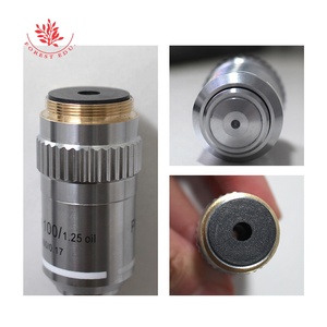
Oil Immersion
The 100x lens uses an oil immersion technique to improve resolution and focusing. This technique reduces light refraction by using a special type of oil between the lens and slide. The result is a clearer, more detailed image, especially for small specimens. It is essential in fields that require seeing fine cellular details, such as pathology. The improved clarity makes it a standard tool for examining thin tissue sections.
-
Clinical Microscopy
This objective is widely used in clinical microscopy, particularly in medical laboratories. It allows pathologists to study tissue samples with great detail. The high level of detail supports accurate diagnoses in medical practices. In this context, seeing small cancer cells or tissue structures can be vital for patient treatment. Its clarity and precision make it indispensable in disease diagnosis and research.
-
Fluorescence Microscopy
The 100x objective is compatible with fluorescence microscopy, allowing scientists to study marked cells and tissues. This technique lets researchers track specific components in live cells, offering dynamic biological insights. Fluorescence microscopy is commonly used in cellular biology and genetics to trace proteins or DNA with great precision. This objective significantly enhances the visualisation capability in these advanced research areas.
-
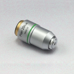
High Numerical Aperture
The 40x objective lens is designed with a high numerical aperture (NA), which allows it to gather more light and resolve finer details. This is particularly useful when observing translucent or small specimens, as it improves image contrast and clarity. A high NA enhances the lens's optical performance, making it suitable for advanced microscopy in biological and material sciences.
-
Wider Application Range
This objective lens is versatile and can be used in various microscopy techniques, including brightfield, phase contrast, and fluorescence. Its adaptability makes it a valuable tool in the laboratory for different specimen preparations. Whether used in a biological study for cell observation or in material science for material testing, the 40x lens accommodates a wide range of needs.
-
Anti-Reflective Coatings
The 10x objective lens features anti-reflective coatings on its surface. These coatings reduce unwanted reflections that can otherwise decrease image brightness and sharpness. By minimising glare, the lens ensures more comfortable and accurate viewing. In microscopy, this means cleaner, clearer images that reveal finer details. The coatings improve efficiency by allowing more light to pass through to the specimen.
-
Phase Contrast
The 4x objective is especially useful for observing unstained, transparent specimens through phase-contrast microscopy. This technique enhances contrast without staining, making it ideal for live cells or thin tissues. It allows users to view specimens in their natural state, vital for biological studies. The lens improves visibility of cell structures, preserving specimen integrity for dynamic observations.
How to Choose Microscope Objectives
-
Application Requirements
Considering the specific needs of the intended application is crucial. For instance, biology and medicine may require objectives that offer high resolution and precise detail for cellular studies. Material science, on the other hand, might focus on objectives that provide clear views of metals or polymers. Therefore, identifying the purpose of the microscopy is critical in choosing the right objective lens.
-
Specimen Type
The type of specimen being examined greatly influences objective selection. Living cells, tissues, or small organisms typically necessitate objectives tailored for biological microscopy. These should be highly resolving and often use techniques such as oil immersion or phase contrast. For materials like plastics and metals, objectives that offer clear, detailed images in brightfield or polarized light are more suitable.
-
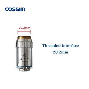
Objective Compatibility
The chosen objectives should fit within the microscope specifications. These include the correct mounting thread and the right type of connections for the eyepiece. It is also necessary to ascertain if the microscope supports different types of objectives, such as those for oil immersion or phase contrast. This guarantees that there is flexibility for future needs and upgrades.
-
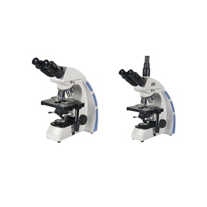
Microscope Type
The type of microscope in question determines the objective lenses that can be used. Standard light microscopes use brightfield objectives to view specimens under normal illumination. Meanwhile, fluorescence or confocal microscopes require specialized objectives designed for viewing fluorescently marked samples. Identifying the microscope type helps in selecting compatible objectives that maximise the equipment's potential.
-
Budget Considerations
While high-quality objectives are crucial for precise microscopy, they can be costly. Thus, it's essential to weigh the expenses against the benefits for specific applications. Sometimes, a mid-range objective can meet typical lab needs. In other cases, a high-end objective is necessary. Careful evaluation helps make an informed decision without overspending.
-
Performance Characteristics
Key performance traits include magnification power, numerical aperture, and resolving ability. A high numerical aperture enables the objective to gather more light, improving resolution and image quality. Meanwhile, resolving power indicates the smallest detail the lens can distinguish. It affects clarity, particularly in fine-structured specimens. These factors determine the objective's effectiveness for the intended microscopy application.
Enhancing Microscope Objectives
Durability
-
Proper Handling
Great care should be exercised when handling microscope objectives. This is because careless handling can lead to scratches on the lens or damage to the mounting. When changing objectives, one should do it slowly and always handle them by the side, never touching the glass. Moreover, gently placing them in designated holders when not in use minimises the likelihood of such damages occurring.
-
Regular Cleaning
Microscope objectives should be cleaned regularly using suitable techniques and materials. Improper cleaning can result in scratches or damage to the lens surface. One is advised to use only approved lens paper or microfiber cloth along with recommended lens cleaning solutions. Never using abrasive materials or harsh chemicals that may damage the coating. This ensures that the objectives have a long life.
-
Avoid Direct Contact
Using an oil or immersion lens remotes the objective from direct contact with the specimen. This not only protects the lens but also provides clearer images. Users should avoid placing any solid materials directly on the lens surface. Doing so will ensure the long-term wear and tear of the microscope.
-
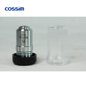
Use Lens Caps
Always using lens caps after removing objectives from the microscope protects them from dust and scratches. Dust can settle on the lens surface over time, leading to a decrease in image quality. Similarly, scratches on the lens will result in lower image quality. Lens caps are essential in prolonging the life of these important tools.
-
Storage Conditions
Store objectives in a dry, controlled environment. Exposure to humidity and extreme temperatures can damage the lens coatings. In addition, high humidity can also promote fungus growth which will damage the objective. Thus, a sturdy, protective case maintains microscope conditions and integrity.
-
Routine Inspections
Routine inspections for signs of wear help address minor issues before they cause major damage. Additionally, checking for cracks or chips in the lens will help maintain image quality. Catching early signs of damage before they become major problems will also help save costs and enhance performance. Regular objective maintenance will ensure optimum functionality.
Q & A
Q. What does a high numerical aperture (NA) mean for an objective lens?
A. A high numerical aperture improves the lens's ability to gather light. This gives sharper and more detailed images, especially of small or fine specimens. It also enhances contrast, making it more useful for viewing transparent or unstained samples. High NA objective lenses are especially valuable in advanced microscopy, enhancing optical performance.
Q. Why is routine cleaning important for microscope objectives?
Q. It is important to clean microscope objectives frequently as contaminants like dust or oil residue decrease image quality. Furthermore, improper cleaning methods lead to lens scratches or damage. Therefore, regular cleaning ensures optimal performance. Also, using appropriate tools and materials when cleaning prevents future damage to the objectives.
Q. What is the purpose of an oil immersion objective?
O. It increases light-gathering capability by filling the space between the lens and the slide with oil. This improves resolution and magnification for viewing small specimens. The oil has a similar refractive index to the lens, reducing light refraction and enhancing image clarity. This is crucial for detailed cellular structures in biological microscopy.
Q. How do objective lenses in microscopy differ from those in other optical instruments?
Microscope objectives are designed to provide more power and focus for tiny specimens. This is because these specimens are usually in the range of a few micrometres. On the other hand, telescope objectives are designed to magnify distant objects like stars and planets. This is more than microscope objectives. Moreover, microscope objectives use oil immersion, which improves detail for small biological specimens. Telescope objectives do not use this method for large cosmic objects.
Q. What factors should be considered when selecting a microscope objective?
Specimen type, intended application, and the compatibility of the existing microscope objectives are among the factors to consider. Also, the viewing medium, such as air or oil, and the magnitude of the refractive index of the medium must be considered. This is because they should all match. These are basically the key factors to consider when choosing microscope objectives.


 浙公网安备 33010002000092号
 浙B2-20120091-4